
26 August 2025
By Ankita Thawani, Travelling Fellowship (Development) recipient
Ever wonder how you can follow the smell of fresh pastries to the break room, dodge a ball flying at your face or get a reflexive jump scare when your friend yells “Boo!”? The answer lies between your ears, literally! We vertebrates have a complex head that is a nexus of sophisticated sensory organs and a highly tuned central nervous system (CNS), giving us an evolutionary advantage for both predation and survival. In contrast, our closest living non-vertebrate cousins, the tunicates (also known as sea squirts), stay glued to rocks, filter-feeding their lives away.
But let’s not get too smug. Tunicates do have sensory organs and a brain, though much simpler than ours. In fact, the gene signatures in the embryonic progenitor cells that develop into sensory organs are strikingly similar between vertebrates and tunicates. So, what led to the evolution of our much more complex heads?
I am a developmental biologist specializing in early vertebrate embryogenesis, using the mouse as a model organism. Last summer, I began a collaboration with Alberto Stolfi and his lab at Georgia Institute of Technology in Atlanta, USA, thanks to a Travelling Fellowship from Development, supported by funding from The Company of Biologists. I spent two weeks in the Stolfi lab learning tunicate embryology, equipping myself with the technical expertise needed to compare vertebrate and non-vertebrate embryonic development. As an advanced postdoctoral researcher seeking faculty positions, my long-term goal is to understand the genetic basis of vertebrate head evolution.
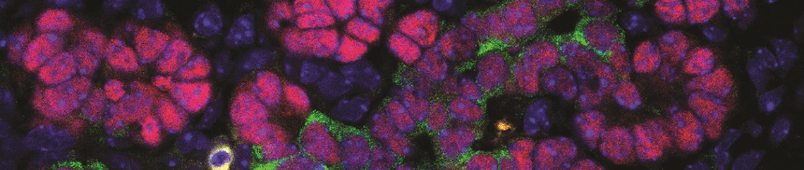
Before my research visit, I identified several transcription factors important for vertebrate craniofacial patterning, including some forkhead box transcription factors. In vertebrates, these genes are associated with cranial sensory placode cell fate specification (which gives rise to sensory organs), sensory organ development, neural tube induction and more. Interestingly, expression of orthologs of these genes can be found at comparable stages of embryonic development in tunicates, as shown in gene expression databases. Comparing the function of the orthologous gene across the vertebrates and tunicates (non-vertebrate chordates) is an important question to understand conservation of gene regulatory networks across the phylum.
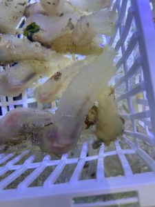
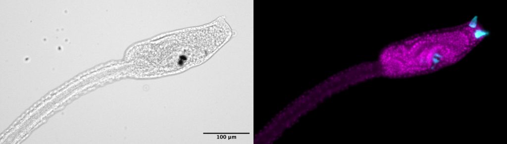
The Stolfi lab works with the tunicate model organism Ciona robusta (formerly Ciona intestinalis type A) to understand the molecular mechanisms of neurodevelopment in chordates. In the Stolfi lab, I gained technical knowledge of embryonic tunicate aquaculture—identifying and handling gravid adult animals, harvesting gametes, performing synchronous ex vivo fertilization, harvesting embryos at desired developmental stages and electroporating tunicate embryos to deliver reporters and loss-of-function constructs. These animals undergo embryonic development from a single cell zygote to a swimming larva within 17 hours post-fertilization, making this animal model high throughput.
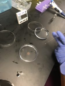
To electroporate tunicate embryos, it is essential to remove the chorion, a protective layer of extracellular material and follicle cells that will protect the embryo proper inside. This is one of the most technically challenging steps of tunicate embryology. I worked with Chris Johnson and Sydney Popsuj in the Stolfi lab to learn multiple methods of gamete harvesting, fertilization, and dechorionation of tunicate embryos. In addition, I worked with Chris to knock out one of the candidate craniofacial development genes I had previously identified at the one-cell stage in tunicate embryos using CRISPR-Cas9 technology. Chris had generated small guide RNAs (sgRNAs) designed to knock out the gene of interest. We tested these sgRNAs in vivo by electroporating a plasmid concoction including a ubiquitously expressed Cas9 and evaluated the larval morphology. We also added reporter constructs to the plasmid mix to: a) observe the success of electroporation via fluorescent reporters, and b) observe cell lineages in control versus crispants (animals which are recipients of CRISPR-Cas9).
During those two weeks of training, I presented a seminar to the Stolfi lab about my published postdoctoral work and ideas for my independent research program. At the end of my training, I shipped electroporated and fixed embryos to my postdoctoral laboratory (Andy Groves’ lab at Baylor College of Medicine, Houston, USA) for high-resolution microscopy. I also collected embryos between 4 hpf and 7 hpf, a time range when we believe progenitors for tunicate sensory organs and brain are specified and prepared them for RNA histology with Sydney’s help to investigate the spatio-temporal expression of the transcription factors of interest in tunicate embryogenesis.
Since my research visit, I established these techniques in-house in my postdoctoral lab, and we now have our own aquarium and electroporation setup to continue this project. Furthermore, I am currently training a graduate student in the Groves lab to implement these techniques for their project. This training visit was the start of a sustainable collaboration with the Stolfi lab that will continue with my career transition as an independent investigator. The expertise of the Stolfi lab in tunicate and molecular biology and my experience in vertebrate embryology and histological techniques will help us answer some fundamental questions of development and evolution in vertebrates.
Note: Ankita Thawani and the Groves laboratory research group have moved to Washington University in St. Louis – School of Medicine, USA, as of May 2025.
You must be logged in to post a comment.